Scaffolding Protein’s L-Shaped Conformation Revealed
When CSSB group leader Christian Löw and his colleagues were unable to crystallize the scaffolding protein, PDZK1, they did not give up the hope of uncovering detailed structural information about this important protein. Löw turned to his EMBL colleague Dmitri Svergun, who runs the P12 beamline at PETRA III, for his help in performing a systematic study of PDZK1 using small-angle X-ray scattering (SAXS). Loew and Svergun’s collaboration not only resulted in the publishing of a structural model of PDZK1 in the journal “Structure” but also demonstrates the power of using an integrated approach to solve complex structural biology questions.
The Unattainable Crystal
Similar to metal scaffolding which provides construction workers with access points to a building, scaffolding proteins mediate interactions between proteins situated on the membrane of the human cell. The scaffolding protein PDZK1 is comprised of four PDZ domains, three linkers and a C-terminal tail. The molecular structure of each of the four individual PDZ domains has been solved using X-ray crystallography and NMR spectroscopy, however the overall arrangement of the domains in the protein as well as their interactions was not yet understood.
The individual PDZ domains of PDZK1 are small and highly conserved making them straightforward to crystallize and thus ideal for x-ray crystallography experiments. However, despite numerous attempts the Löw group was unable to generate a crystal of the entire PDZK1 protein. “With PDZK1 we realized that we would need to a use a different method to gain more structural data; a method that does not require the crystallization of the sample,” explains Löw.
Revealing a Defined Shape
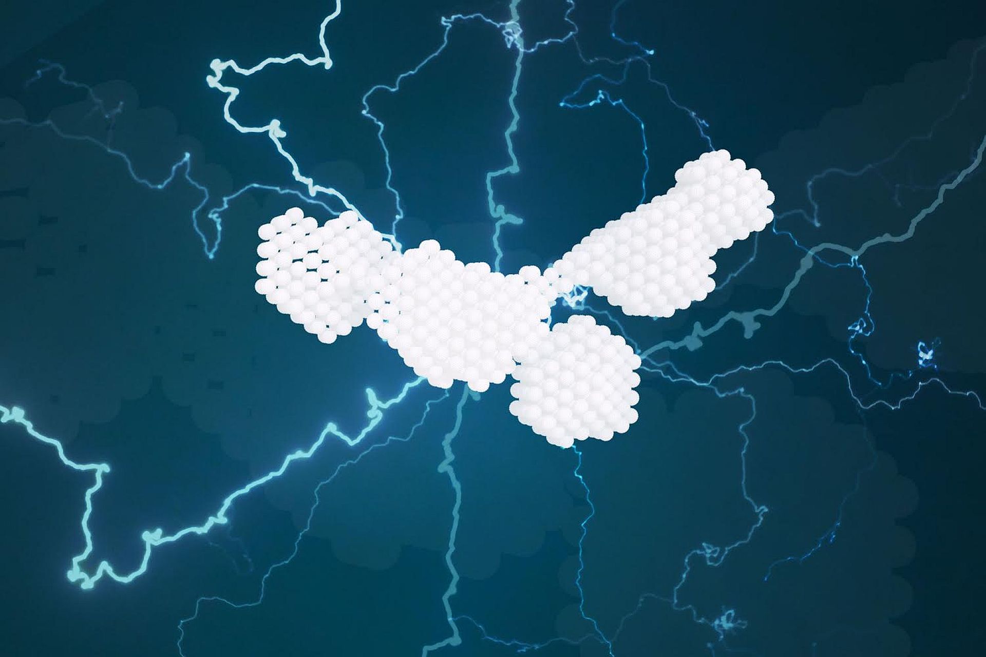
Small-angle X-ray scattering (SAXS) is an x-ray based technique that allows the analysis of proteins and other molecules in solution. “SAXS is a powerful method to obtain shape information of molecules,” explains Svergun. Together, the Löw and Svergun groups performed a systematic study using SAXS experiments and determined a solution structure of PDZK1 that held some unexpected surprises.
Bioinformatics tools had suggested that PDZK1’s PDZ domains and linkers would behave like beads on a string moving around in a highly flexible manner. The SAXS experiments revealed however, that PDZK1 has a relatively defined L-shaped conformation with only moderate flexibility. The experiments also revealed that PDZK1 linker regions are not simply disordered spacers but actively contribute to the proteins L-shaped confirmation. “Taking a closer look at these intermediate regions will ultimately help us to fully understand the function and dynamics of PDZK1 as well that of other scaffolding proteins,” notes Nelly Hajizadeh, one of the first authors of the study.
For the future, Löw and his group plan to examine PDZK1 in complex with binding partner using electron cryo-microscopy. Cryo-EM is similar to SAXS in that it does not require a crystallized sample however the resulting images generated by cryo-EM can achieve higher resolution and reveal more structural detail. Löw is hoping to use the new cryo-EM facility in the basement of the CSSB building for the next step in this project. “Only by integrating a range of techniques and collaborating with colleagues, will we be able to fully understand complex proteins like PDZK1,” states Löw.
Original Publication:
Probing the Architecture of a Multi-PDZ Domain Protein: Structure of PDZK1 in Solution; Nelly R. Hajizadeh, Joanna Pieprzyk, Petr Skopintsev, Ali Flayhan, Dmitri I. Svergun, Christian Löw; Structure, 2018; DOI: 10.1016/j.str.2018.07.016