Missing link in replication process of coronaviruses identified
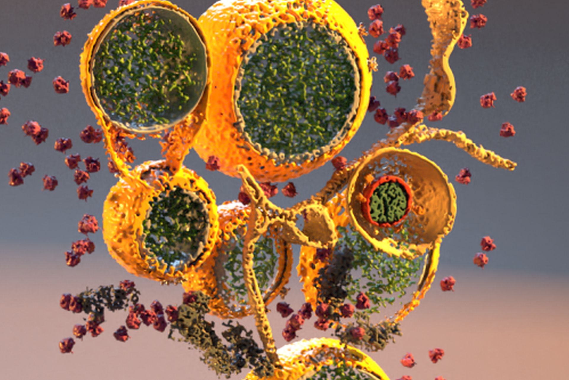
Coronaviruses such as SARS-CoV-2 drastically remodel infected cells by building special membrane structures that accommodate virus replication. It is already known that the genome of the virus is copied in these replication organelles. These structures seemed to be completely sealed, which made it puzzling how the newly made genomes could leave this compartment. In cooperation with scientist from the Grünewald (HPI, UHH) group, researchers from Leiden University Medical Center now found a passage mechanism that is mediated by a large crown-shaped molecular pore. This protein structure is a potential new starting point for the development of antiviral drugs. The study has just been published online in the renowned journal Science.
A research team from the Leiden University Medical Center (The Netherlands, LUMC), in cooperation with the Hamburg-based Research Department “Structural Cell Biology of Viruses” at Centre at Structural Systems Biology (CSSB) and the Heinrich Pette Institute, Leibniz Institute for Experimental Virology (HPI), has been working together to focus on the 'remodeling' and damage caused by the replicating coronavirus in the infected cell.
Virus builds special compartment for genome replication
Coronaviruses convert membrane structures of infected cells into apparently closed compartments in which the viral genetic material, the RNA, is copied. These ‘replication organelles’ are surrounded by a double membrane layer, which probably offers optimal conditions for the viral genome copying process. However, the replication organelles may also allow the virus machinery to hide from certain cellular immune responses.
The newly made virus RNA carries the code to make new virus proteins, and ultimately has to be packaged in new virus particles in order for the virus to spread. This requires the export of the newly made RNA from the replication organelles. Exactly how this export occurs was however unknown.
Electron cryo-microscopy
In the recently published study, the research team used electron cryo-tomography to analyze the replication organelles of the coronavirus, a technique that enables cell structures to be viewed with high resolution in their native context. As a result, the research team identified an opening in the double membrane of the replication organelle: a combination of viral proteins creates a pore that might enable the export of RNA. “Tomograms of infected cells allowed us to retrieve the structure of the molecular pore at nanometer resolution,” explains Dr. Ulrike Laugks, a member of the Grünewald group “part of the data including those on SARS-CoV2, was recorded and gathered at the cryo-EM facility at CSSB, which is strongly supporting scientists during these extraordinary times.”
New starting point for the development of virus inhibitors
“This newly discovered connection between the replication organelle and the rest of the cell not only provides more insight into the organization of the replication process of coronaviruses, but also offers a new starting point for the development of antiviral drugs”, explains LUMC researcher Prof. Eric Snijder. Blocking this pore is likely to inhibit or arrest the multiplication of coronaviruses.
With this discovery, the research team found one of the missing pieces in the puzzle of coronavirus replication. Follow-up research will have to show how the newly discovered structure functions and whether it is indeed a useful target for the development of coronavirus inhibitors.
Reference:
Wolff G. et al. (2020) A molecular pore spans the double membrane of the coronavirus replication organelle. Science
https://science.sciencemag.org/content/early/2020/08/05/science.abd3629