Super-resolution fluorescence microscopy has revolutionized biological imaging. However, resolution improvement is only one side of the story. Equally important is structural preservation of the sample. We are combining fast-freezing techniques (vitrification) with the concept of super-resolution imaging to achieve both.
Super-resolution fluorescence microscopy under cryo-conditions (super-resolution cryo-FM) is a new field of microscopy that is aiming to combine super-resolution concepts with the benefits of cryo-immobilized samples. Structural preservation of the sample is critical for imaging with high resolution to prevent artefacts and misinterpretation of the data. Super-resolution cryo-FM has two main application fields:
1) Provide an alternative to conventional (ambient temperature) super-resolution FM methods, where currently the common practice for immobilization is chemical fixation.
2) Bridge the resolution gap in correlative light and electron cryo-microscopy (cryo-CLEM) to take full advantage of the complementary features of both imaging modalities.
Research Focus
We are working on the intersection of physics, engineering and biology. Key research interests of our group are:
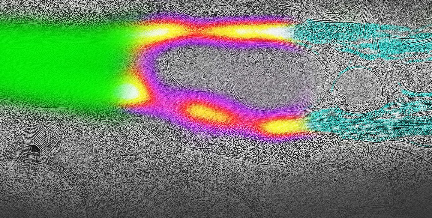
Cryo-single-molecule photo-physics: The key for super-resolution imaging in general is photo-switching of the fluorophores. As this is only poorly studied and understood under cryo-conditions, one of our major aims is to gain a deeper insight into the photo-physical mechanisms of different molecules at a temperature suitable for vitreous biological specimens (devitrification point of water: -135°C).
Cryo-single-molecule photo-blinking. IMAGE: Flackenhayn et al. (2026)
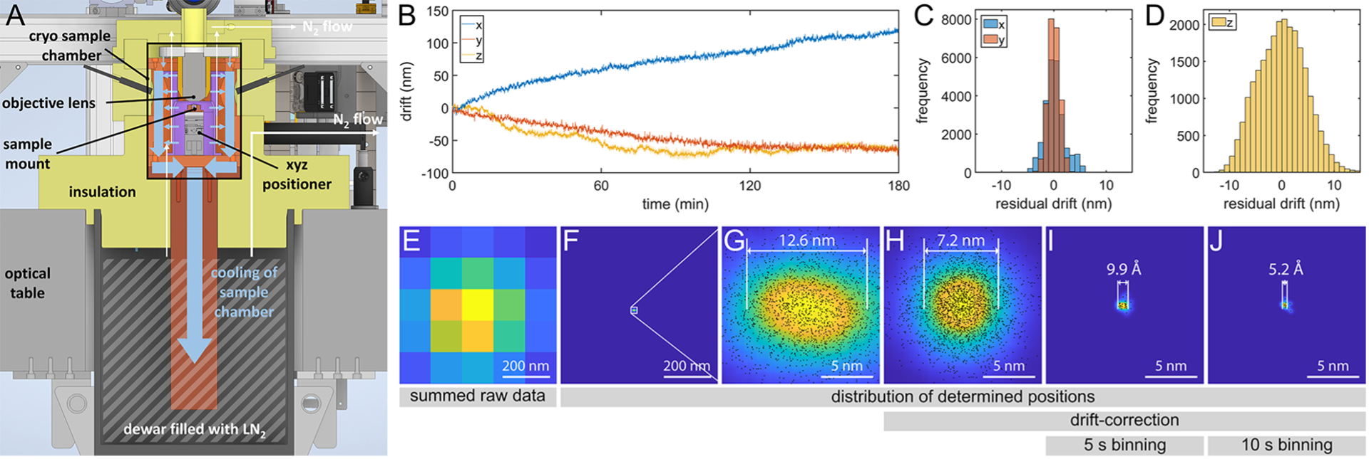
Development of new cryogenic optical imaging systems: We are developing new techniques and methodology with a focus on ultra-high stability systems for single-molecule investigations and super-resolution cryo-FM with a particular interest on super-resolution cryo-CLEM. Minimizing the resolution gap in cryo-CLEM is opening up a broad spectrum of applications in the field of structural cell biology.
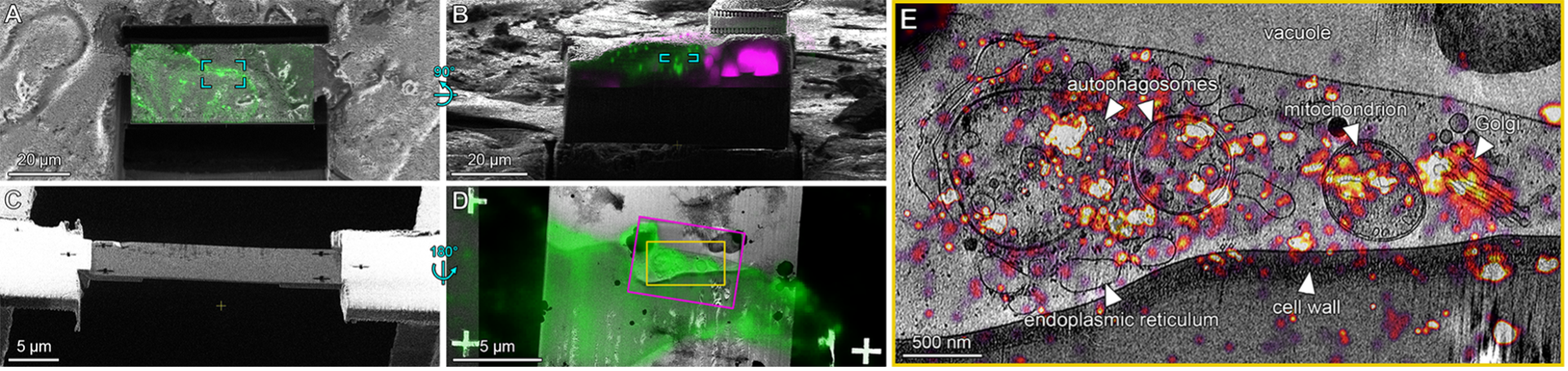
Application of super-resolution cryo-CLEM in the field of structural cell biology: In close collaboration with other groups in the CSSB and beyond, we are using super-resolution cryo-CLEM to tackle current biological questions in the context of infectious diseases, translation mechanism or nuclear architecture. Integrating cryo super-resolution fluorescence microscopy into the cryo-EM workflow enables for example identification and localization of rare biological events in host-pathogen interactions.
Super-resolution cryo-CLEM methods developed in the Kaufmann lab
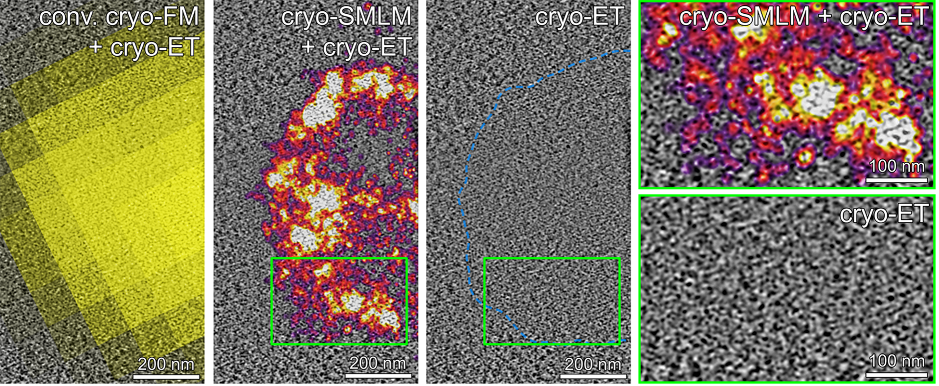
Cryo-SMLM: Cryogenic single molecule localization microscopy (cryo-SMLM) utilizes fluorescent on-states in the seconds range to localize individual molecules with a precision in the sub-10-nm range.
Enables cryo-CLEM on the single-molecule level.
Allows visualizing the distribution of specific proteins in the structural context of cryo-ET with single-digit-nm precision.
Many common FPs (e.g. EGFP, EYFP, mCherry) and organic dyes (e.g. JF525, Atto647) are suitable for cryo-SMLM.
Compatible with any cryo-ET workflow, incl. advanced procedures such as cryo-lift-out.
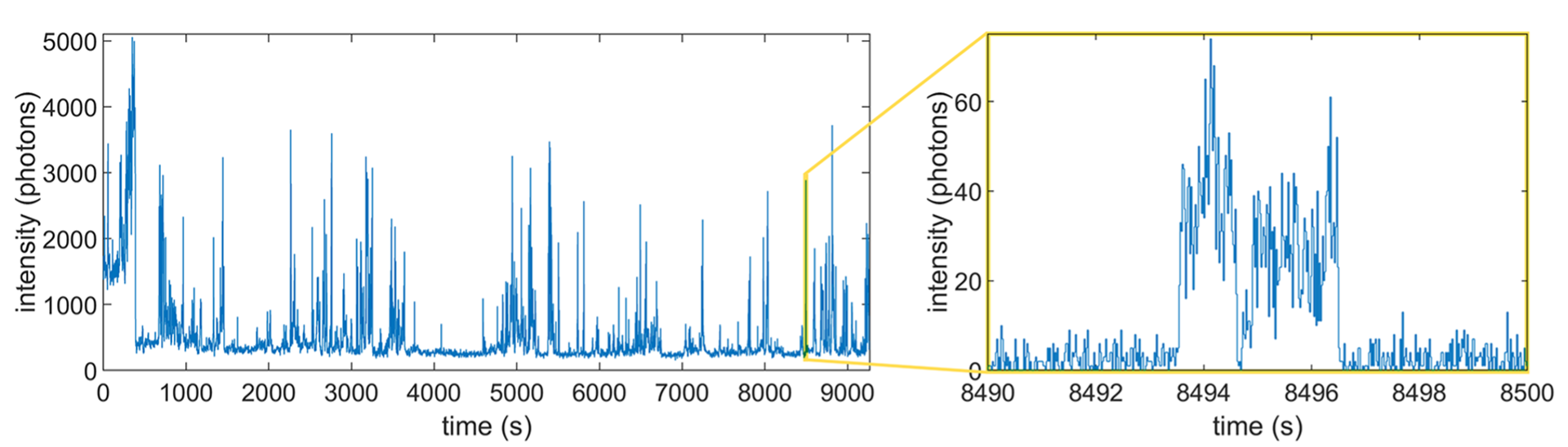
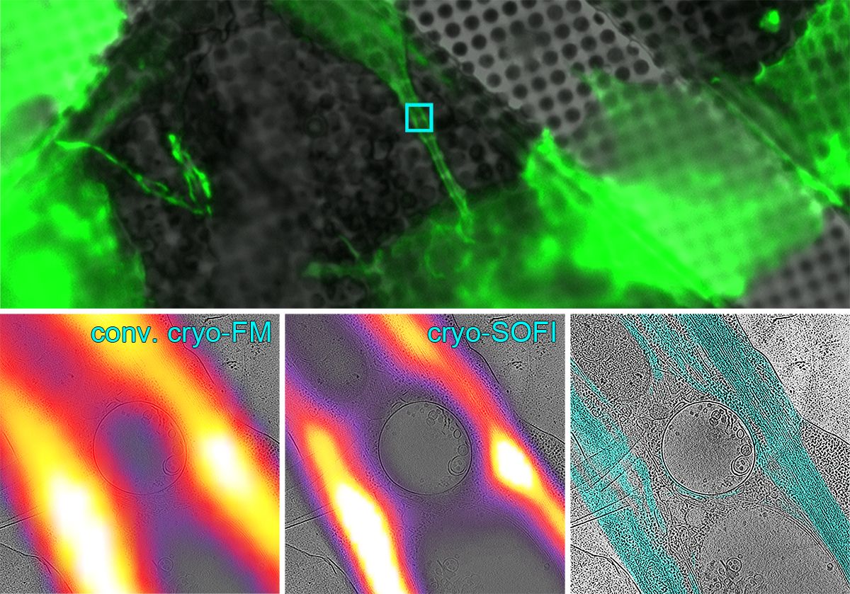
Cryo-SOFI: Cryogenic super-resolution optical fluctuation imaging (cryo-SOFI) utilizes fast cryo-photo-blinking of fluorescent molecules to increase resolution in cryo-FM.
Operates with low laser intensities and achieves a resolution enhancement of 2-3x.
Achieves single molecule sensitivity and elimination of non-blinking background.
High versatility: Works with almost all fluorescent proteins and organic dye molecules.
High efficiency: Data acquisition typically requires only 2 min.
Correlative cryo-SOFI and cryo-ET. IMAGE: Moser & Pražák et al. (2019)
Future projects and goals
Super-resolution cryo-FM/cryo-CLEM is still at an early and experimental stage. Overcoming technical challenges and gaining a better understanding of the underlying photo-physics will help us to push the boundaries of this method and turn it into a powerful tool for structural cell biology.
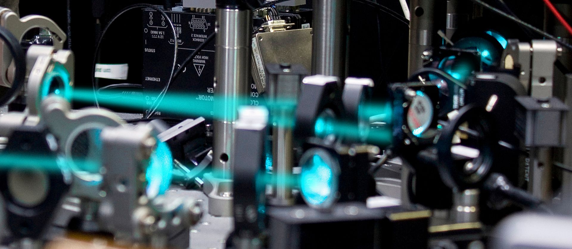
Microscope setup on optical bench. IMAGE: R. Kaufmann.
Current available BSc and MSc projects
Listed projects are aimed at Physics or Nano Science students, but open to other disciplines too. If you are interested, simply contact us for more information.
Listed projects are aimed at Physics or Nano Science students, but open to other disciplines too. If you are interested, simply contact us for more information.
Falckenhayn J, Duong VQ, Prabhakar N, Harley I, Yuen ELH, Bozkurt TO, Carter SD, Pražák V, Kaufmann R (2026) On-lamella super-resolution cryo-CLEM for cryo-ET enabled by vacuum-free ultra-stable cryogenic fluorescence microscopy. bioRxiv. 717675; https://doi.org/10.64898/2026.04.14.717675
Gebauer R, Machala EA, Mironova Y, Jönsson MR, Mazur J, Feldmann CA, Zimmeck MA, Silvester E, Caragliano E, Falckenhayn J, Yuen ELH, Ibrahim T, Hellert J, Bozkurt TO, Kaufmann R, Quemin ERJ, Grünewald K, Pražák V (2026) View Tomo: Context-aware targeting and analysis in electron cryo-tomography.bioRxiv.719727; https://doi.org/10.64898/2026.04.21.719727
Pražák V, Harley I, Falckenhayn J, Boutell C, Thomason PA, Davis BG, Kaufmann R, Carter SD (2026) In situ molecular architecture of PML bodies reveals open-state columnar trinucleosome assemblies within a porous, chromatin-permissive interior. bioRxiv 2026.06.07.729032; doi: https://doi.org/10.64898/2026.06.07.729032
Gartenmann L, Wainman A, Qurashi M, Kaufmann R, Schubert S, Raff JW, Dobbie IM (2017) A combined 3D-SIM/SMLM approach allows centriole proteins to be localized with a precision of∼ 4–5 nm. Current Biology 27: R1054-R1055 doi: 10.1016/j.cub.2017.08.009
Johnson E, Kaufmann R (2017) Correlative In-Resin Super-Resolution Fluorescence and Electron Microscopy of Cultured Cells. Super-Resolution Microscopy. Springer, 163-177 doi: /10.1007/978-1-4939-7265-4_14
2016
Wolff G, Hagen C, Grünewald K, Kaufmann R (2016) Towards correlative super‐resolution fluorescence and electron cryo‐microscopy. Biology of the Cell 108: 245-258 doi: 10.1111/boc.201600008
Kong Y, Janssen BJ, Malinauskas T, Vangoor VR, Coles CH, Kaufmann R, Ni T, Gilbert RJ, Padilla-Parra S, Pasterkamp RJ (2016) Structural Basis for Plexin Activation and Regulation. Neuron 91: 548-560 doi: 10.1016/j.neuron.2016.06.018
Wegel E, Göhler A, Lagerholm BC, Wainman A, Uphoff S, Kaufmann R, Dobbie IM (2016) Imaging cellular structures in super-resolution with SIM, STED and Localisation Microscopy: A practical comparison. Scientific Reports 6: 27290 doi: 10.1038/srep27290
2015
Ball G, Demmerle J, Kaufmann R, Davis I, Dobbie IM, Schermelleh L (2015) SIMcheck: a Toolbox for Successful Super-resolution Structured Illumination Microscopy. Scientific reports 5: 15915 doi: 10.1038/srep15915
Zhang Y, Máté G, Müller P, Hillebrandt S, Krufczik M, Bach M, Kaufmann R, Hausmann M, Heermann DW (2015) Radiation Induced Chromatin Conformation Changes Analysed by Fluorescent Localization Microscopy, Statistical Physics, and Graph Theory. PloS one 10: e0128555 doi: 10.1371/journal.pone.0128555
Johnson E, Seiradake E, Jones EY, Davis I, Grünewald K, Kaufmann R (2015) Correlative in-resin super-resolution and electron microscopy using standard fluorescent proteins. Scientific reports 5: 9583 doi: 10.1038/srep09583
2014
Schellenberger P, Kaufmann R, Siebert CA, Hagen C, Wodrich H, Grünewald K (2014) High-precision correlative fluorescence and electron cryo microscopy using two independent alignment markers. Ultramicroscopy 143: 41-51 doi: 10.1016/j.ultramic.2013.10.011
Cremer C, Kaufmann R, Gunkel M, Polanski F, Müller P, Dierkes R, Degenhard S, Wege C, Hausmann M, Birk U (2014) Application perspectives of localization microscopy in virology. Histochemistry and cell biology 142: 43-59 doi: 10.1007/s00418-014-1203-4
Müller P, Lemmermann NA, Kaufmann R, Gunkel M, Paech D, Hildenbrand G, Holtappels R, Cremer C, Hausmann M (2014) Spatial distribution and structural arrangement of a murine cytomegalovirus glycoprotein detected by SPDM localization microscopy. Histochemistry and cell biology 142: 61-67 doi: 10.1007/s00418-014-1185-2
Kaufmann R, Hagen C, Grünewald K (2014) Fluorescence cryo-microscopy: current challenges and prospects. Current Opinion in Chemical Biology 20: 86-91 doi: 10.1016/j.cbpa.2014.05.007
Kaufmann R, Schellenberger P, Seiradake E, Dobbie IM, Jones EY, Davis I, Hagen C, Grünewald K (2014) Super-resolution microscopy using standard fluorescent proteins in intact cells under cryo-conditions. Nano Letters 14: 4171-4175 doi: 10.1021/nl501870p
Wang Q, Dierkes R, Kaufmann R, Cremer C (2014) Quantitative analysis of individual hepatocyte growth factor receptor clusters in influenza A virus infected human epithelial cells using localization microscopy. Biochimica et Biophysica Acta (BBA)-Biomembranes 1838: 1191-1198 doi: 10.1016/j.bbamem.2013.12.014
2013
Seiradake E, Schaupp A, del Toro Ruiz D, Kaufmann R, Mitakidis N, Harlos K, Aricescu AR, Klein R, Jones EY.et al. (2013) Structurally encoded intraclass differences in EphA clusters drive distinct cell responses. Nature Structural & Molecular Biology 20 (8): 958-964
2012
Kaufmann R, Gall JG, Cremer C (2012) Superresolution imaging of transcription units on newt lampbrush chromosomes. Chromosome Research 20 (8): 1009-1015
Huber O, Brunner A, MaierP, Kaufmann R, Couraud PO, Cremer C, Fricker G (2012) Localization microscopy (SPDM) reveals clustered formations of P-glycoprotein in a human blood-brain barrier model. Plos One. 7 (9): e44776
Kaufmann R, Piontek J, Grüll F, Kirchgessner M, Rossa J, Blasig I, Cremer C (2012) Visualization and quantitative analysis of reconstituted tight junctions using localization microscopy. Plos One 7 (2): e31128
2011
Cremer C, Kaufmann R, Gunkel M, Pres S, Weiland Y, Ruckeslshausen T, Lemmer P, Geiger F, Degenhard S, Wege C, Lemmermann N, Holtappels R, Strickfaden H, Hausmann M (2011) Superresolution imaging of biological nanostructures by spectral precision distance microscopy. Biotechnology Journal 6 (9): 1037-1051
Grüll F, Kirchgessner M, Kaufmann R, Hausmann M, Kepschull U (2011) Accelerating image analysis for localization microscopy with FPGAs. Field Programmable Logic and Applications 1-5
Kaufmann R, Müller P, Hausmann M, Cremer C (2011) Imaging label-free intracellular structures by localisation microscopy. Micron 42 (4): 348-352
Kaufmann R, Müller P, Hildenbrand G, Hausmann M, Cremer C (2011) Analysis of Her2/neu membrane protein clusters in different types of breast cancer cells using localization microscopy. Journal of Microscopy 242 (1): 46-54
2010
Müller P, Schmitt E, Jacob A, Hoheisel J, Kaufmann R, Cremer C, Hausmann M (2010) COMBO-FISH enables high precision localization microscopy as a prerequisite for nanostructure analysis of genome loci. International Journal of Molecular Sciences 11 (10): 4094-4105
Bohn M, Diesinger P, Kaufmann R, Weiland Y, Müller P, Gunkel M, von Ketteler A, Lemmer P, Hausmann M, Heermann DW, Cremer C (2010) Localization microscopy reveals expression-dependent parameters of chromatin nanostructure. Biophysical Journal 99 (5): 1358-1367
Cremer C, von Ketteler A, Lemmer P, Kaufmann R, Weiland Y, Müller P, Hausmann M, Gunkel M, Ruckelshausen T, Baddeley D, Amberger R (2010) Far-field fluorescence microscopy of cellular structures at molecular optical resolution. Nanoscopy and Multidimensional Optical Fluorescence Microscopy 3.1‑3.35
2009
Lemme, P, Gunkel M, Weiland Y., Müller P, Baddeley D, Kaufmann R, Urich A, Eipel H, Amberger R, Hausmann M, Cremer C (2009) Using conventional fluorescent markers for far‐field fluorescence localization nanoscopy allows resolution in the 10‐nm range. Journal of Microscopy 235 (2): 163-171
Gunkel M, Erdel F, Rippe K, Lemmer P, Kaufmann R, Hörmann C, Amberger R, Cremer C (2009) Dual color localization microscopy of cellular nanostructures. Biotechnology Journal 4 (6): 927-938
Kaufmann R, Lemmer P, Gunkel M, Weiland Y, Müller P, Hausmann M, Baddely D, Amberger R, Cremer C (2009) SPDM: single molecule superresolution of cellular nanostructures. Proceedings of SPIE 7185: 71850J
2008
Lemmer P, Gunkel M, Baddeley D, Kaufmann R, Urich A, Weiland Y, Reymann J, Müller P, Hausmann M, Cremer C (2008) SPDM: light microscopy with single-molecule resolution at the nanoscale. Applied Physics B: Lasers and Optics 93 (1): 1-12
Black Box Science
ABOUT BLACK BOX SCIENCE
A lot of our scientific culture is quite opaque – almost like a black box in some cases.
Particularly for young scientists it is usually quite difficult to get a clear and real/honest insight into the life of a scientist. What is typically communicated are polished results, straightforward careers and an overly idealistic view of science.
Black Box Science is a video-blog-based SciCom project that provides a (self-)critical view into our scientific culture: blackboxscience.org