CSSB Centre for Structural Systems Biology
Suche
Home Research Research Groups Labahn Group

Labahn Group

Structural Biochemistry

Prof. Dr. Jörg Labahn

Group Leader


Home Institute

Forschungszentrum Jülich
www.fz-juelich.de/ics/ics-6/EN/Home

Our research interest is the molecular pathology of proteins relevant in human health. This includes disease mechanisms based on molecular dysfunction as well as the misuse of host proteins by pathogens in infection. The research approach is focused on structural and biochemical methods directed towards the state dependence of molecular interactions.

Research Projects

Gamma-Secretase in Alzheimer Disease

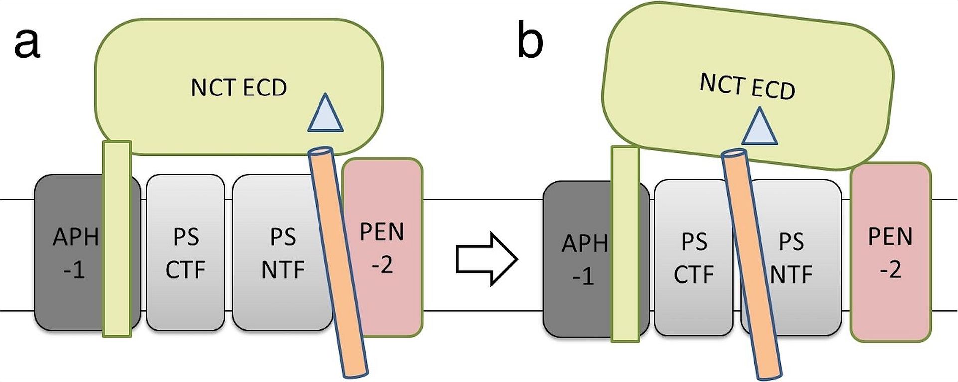
γ- secretase is a membrane protease complex which consists of four subunits: presenilin (PS), nicastrin (NCT), APH-1 and PEN-2 . γ-Secretase cleaves over 90 type-I integral membrane proteins which are involved in many physiological and pathological process.

Uncleaved Presenilin functions als as a Ca-channel whereby it may affect pathologies. Furthermore it is the receptor for papilloma virus infection.

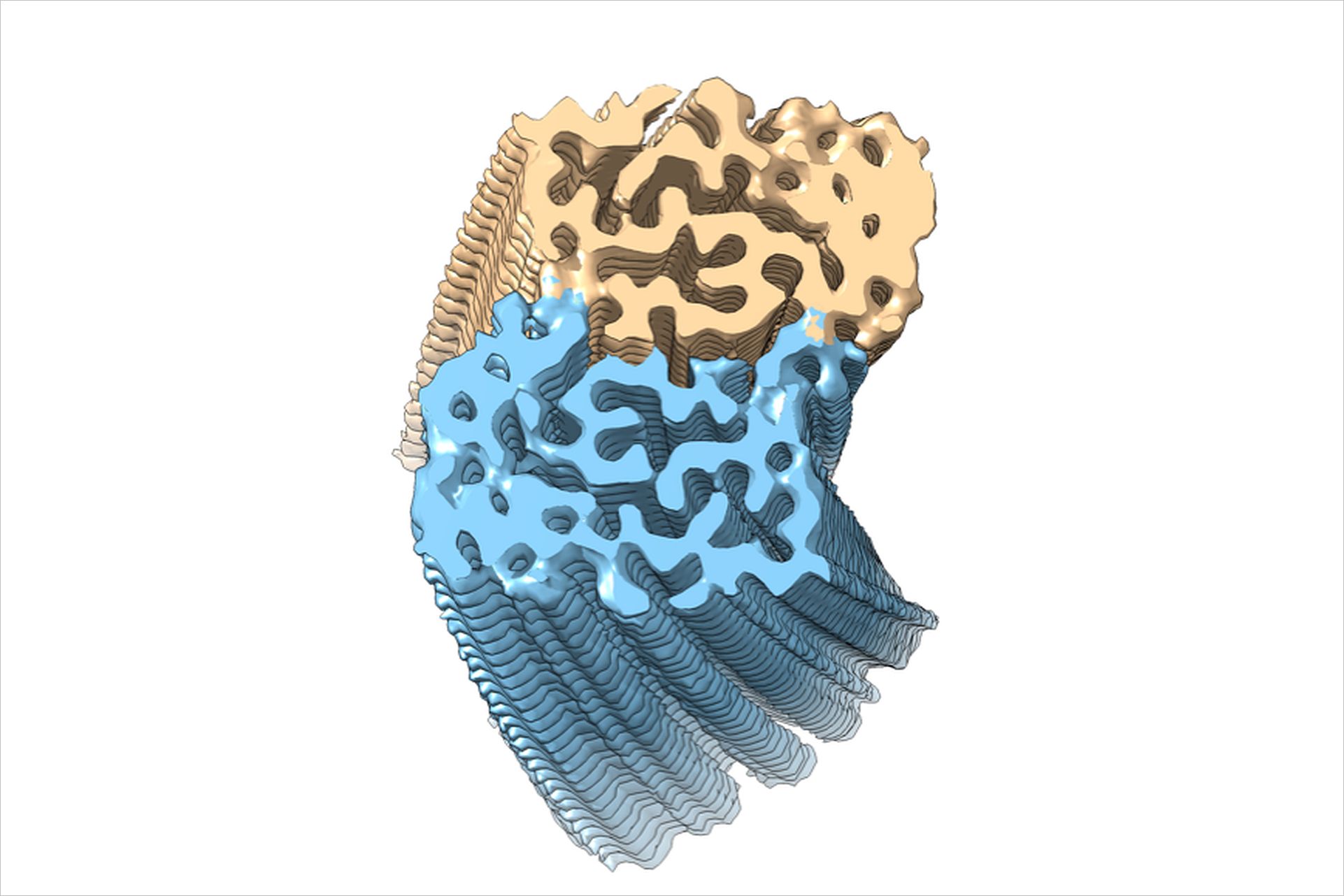
The most widely studied γ-secretase substrates are Notch and C-terminal fragments of β-amyloid precursor protein (APP-CTFs). The cleavage of Notch by γ-secretase releases the intracellular domain to the nucleus, which in turn affects the development of cells and cancer. The cleavage of APP-C99 by γ-secretase generates amyloid-beta peptides (Aβ) of varying lengths, among which the longer peptides -particularly Aβ42 and Aβ43 - form fibrils (s.Fig.1) and are the key components of Aβ plaques, a hallmark of Alzheimer's disease (AD).

We found evidence that NCT (yellow) may is part of an initial substrate docking site(s. Fig. 2)  from where the substrate (orange)  needs to be   translocated to  the activated catalytic subunit PS (grey), where the substrate is processed. The role of APH-1 (dark grey) and PEN-2 (pink) in complex assembly and catalysis is obscure. The pathological change of the product spectrum may be due to a disturbance of substrate translocation.


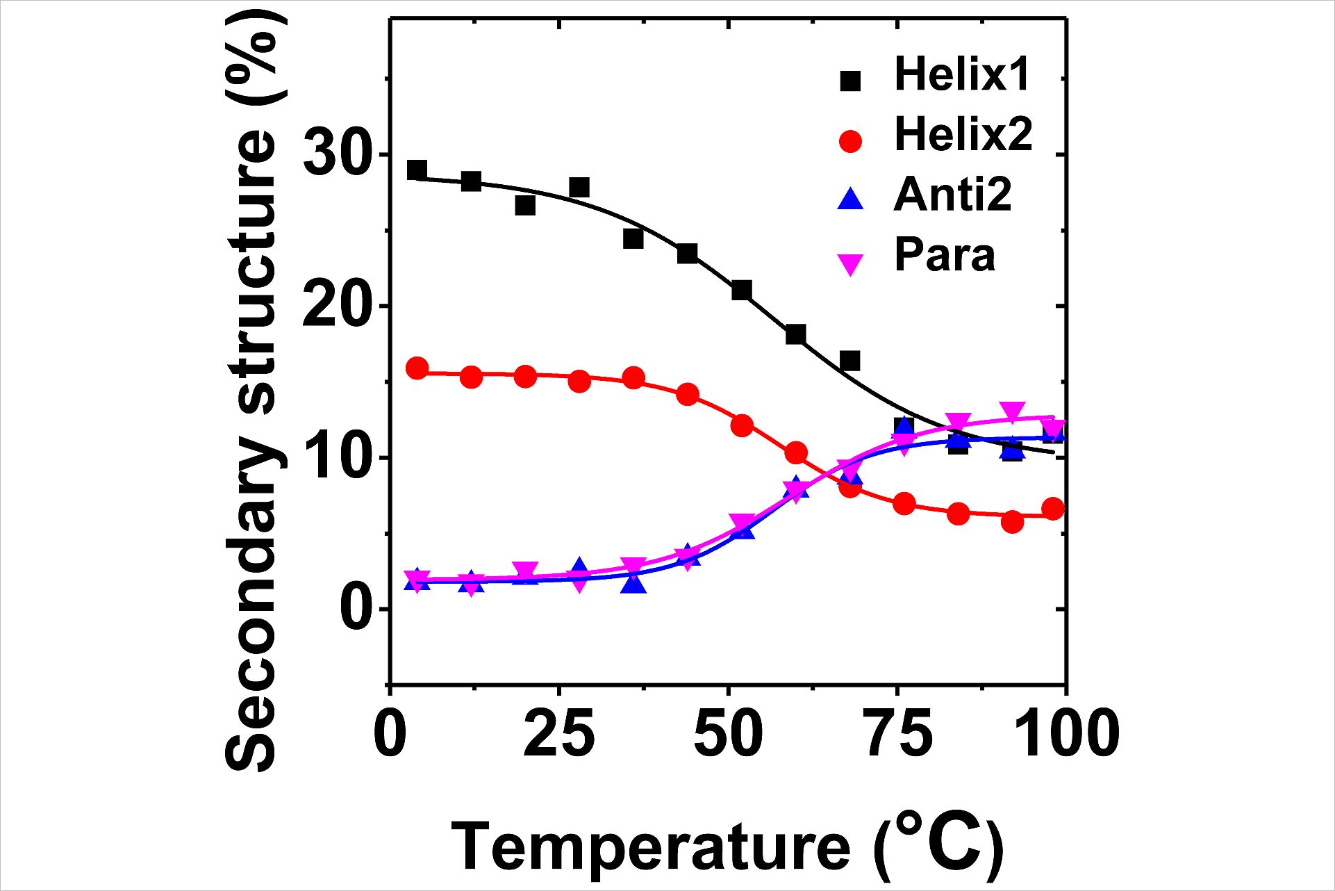
The deconvoluted component spectra of presenilin showed a thermally inducible structural transition from helix to strand (s. Fig. 3). Half of the helical content restructured. Whereas the content of unordered structure and turns increased only slightly, more than ~60% of helical structure turned into β-structure. The linear dependence and the presence of an isodichroic point are evidence for the existence of two different secondary structures of the protein: A low temperature form rich in α-helices and a high-temperature form rich in β-structures. This suggests that the AD related catalytic dysfunction of γ- secretase may be related to structural changes that normally occur only to a small amount at body temperature, but are elevated for mutated Presenilins as found in the cases of familial (inherited) Alzheimer disease.

Par4: Induction of Apoptosis in cancer cells

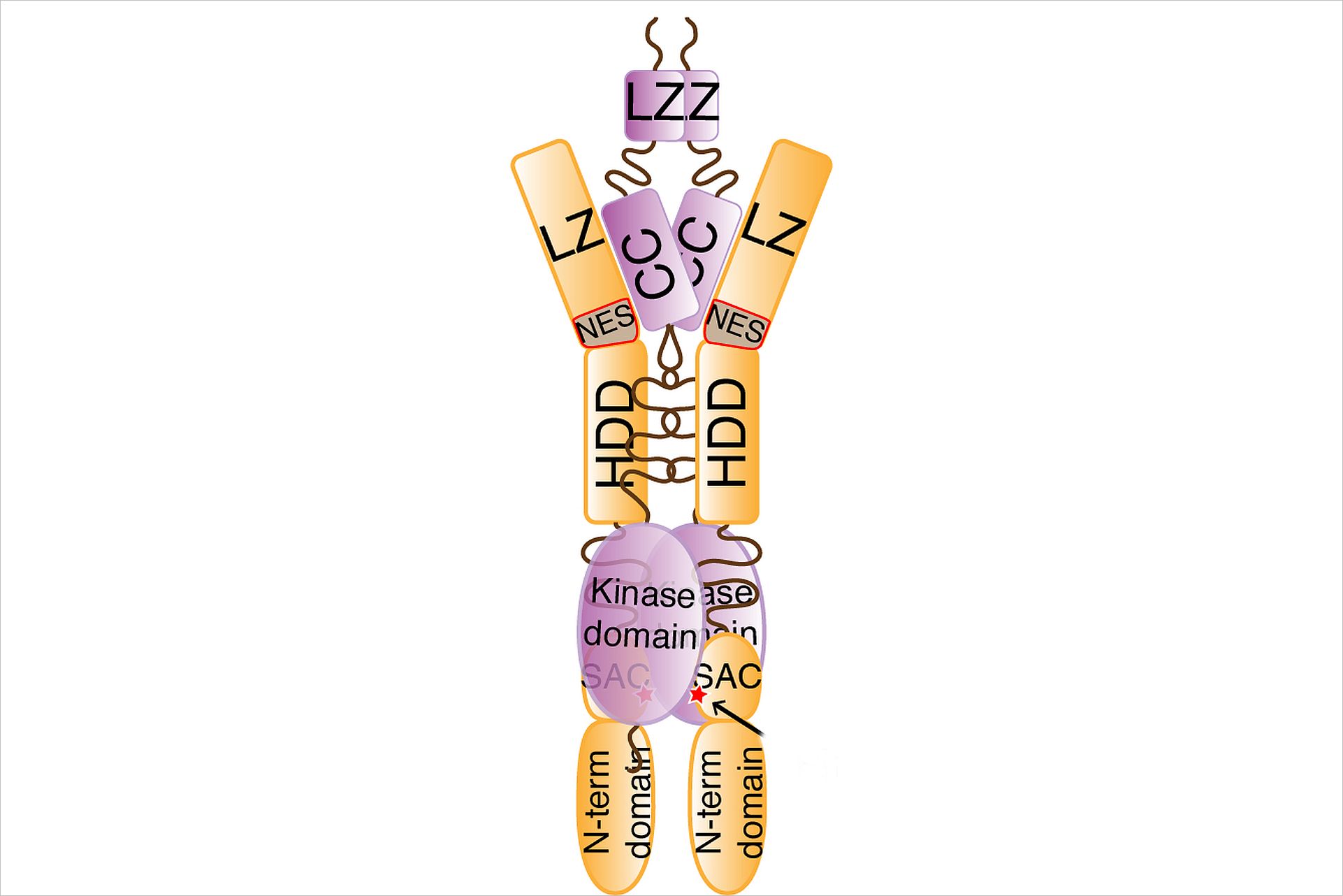
Par-4, the prostate apoptosis response factor 4 (aka Pawr) is a unique pro-apoptotic protein with the ability to induce apoptosis selectively in cancer cells and is involved in HIV-encephalitis. The X-ray crystal structure of the regulatory domain of Par-4 (Par-4CC) showed that Par-4 homodimerizes by forming a parallel coiled-coil structure. The N-terminal half of Par-4CC contains the homodimerization subdomain. This structure includes a nuclear export signal (Par-4NES) sequence, which is masked upon dimerization indicating a potential mechanism for nuclear localization. The heteromeric interaction models specifically showed that charge interaction is an important factor in stability of heteromers of the C-terminal leucine zipper subdomain of Par-4 (Par-4LZ). These heteromer models also displayed NES masking capacity and therefore the ability to influence intracellular localization. An important factor in regulation of the apoptotic function is the formation of a complex with Dapk3 (death associated protein like kinase). The structural information from Par4 allows the formulation of a first model of this complex (s. Fig) and an understanding of the role of the functional domains and subdomains in the regulation of apoptosis.

Membrane Protein Methods

The investigation of membrane proteins in general and mammalian in particular poses substantial problems in obtaining the proteins in sufficient quantity and quality to successfully investigate the mechanisms of their molecular pathology. We developed a lipidic-cubic phase crystallization approach which allows controlling the structure of the meso-phase which provides the crystallization matrix and thereby steering crystallization.


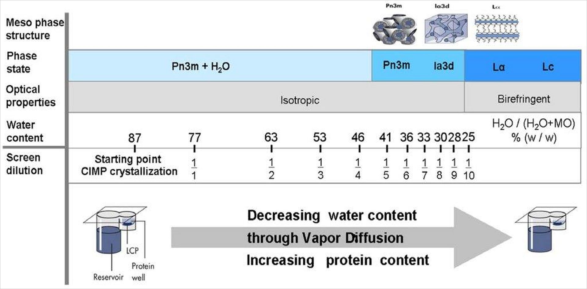
The lipidic meso-phases (Pn3m, Ia3d, Lα, Lc) are formed by monoolein with different amounts of water. The formation of these crystalline phases can be monitored by change of optical properties. Phase formation in the presence of membrane protein leads to insertion of protein into the hydrophobic layers of the monoolein. In case of the cubic phases (Pn3m, Ia3d) 3-dimensional diffusion of the protein within the membrane-like layer is possible: The crystalline meso-phase acts as a solvent for the membrane protein. Reducing the water content of meso-phase by vapor diffusion induces crystallization (phase separation) of the membrane protein. J. Kubicek, et al. PlosOne 7(4): e35458 (2012).

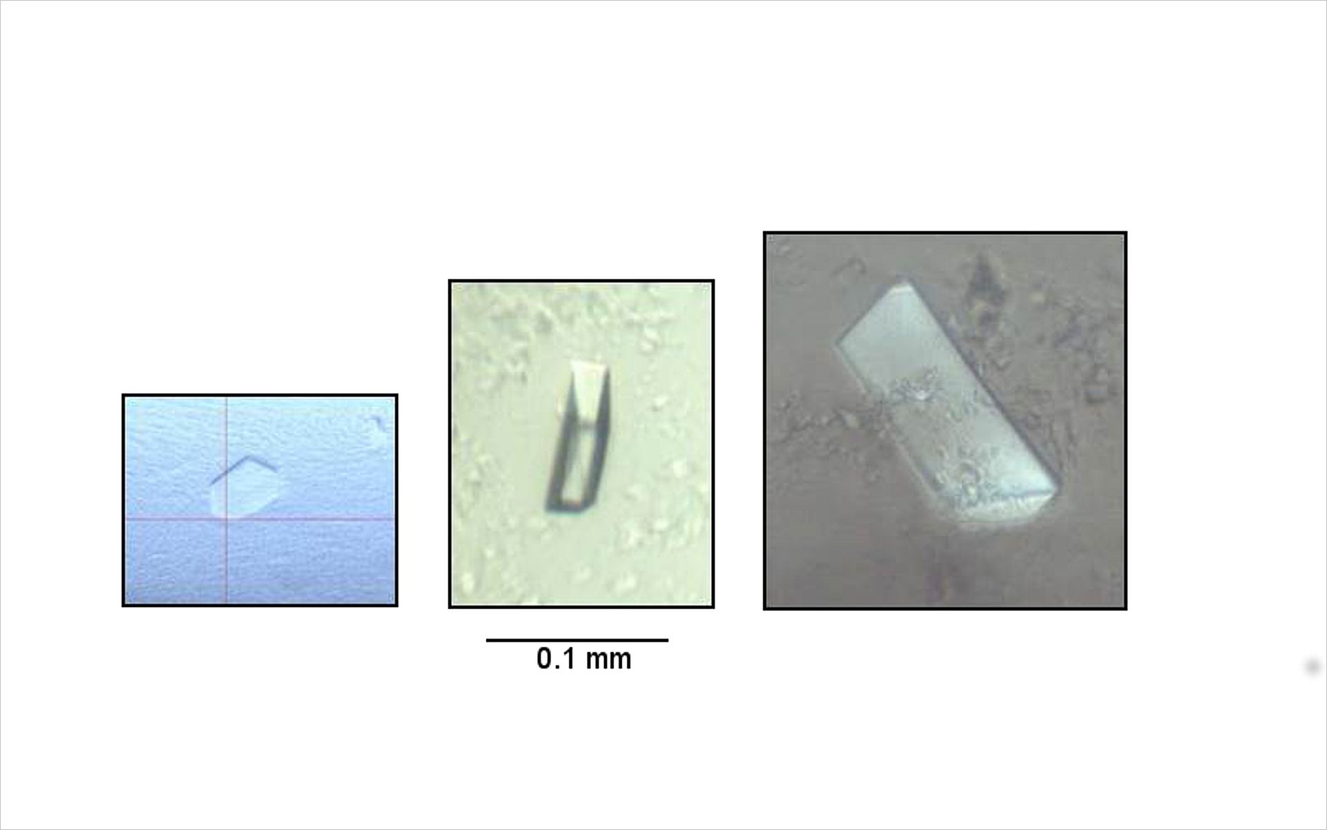
One of the major obstacles in the structural analysis of human membrane proteins is the optimization from a 1st-hit (left) to diffracting crystals (right). If properly optimized crystallization of high quality crystals can be achieved within half a day (see Video)

Controlled In Meso Phase Crystallisation CIMP

The need to solubilize membrane proteins for investigation poses another major problem because detergents can disrupt the protein structure and functionality. An interesting alternative strategy is the solubilisation of membrane proteins by genetic engineering.  20 to 25 % of hydrophobic amino acids at the protein surface need to be mutated to more soluble ones to transform the insoluble membrane protein into a soluble. There is a specific exchange code for aminoacids that minimizes functional and structural disturbances: the QTY code (S. Zhang et al, PNAS, 115 (37) E8652-E8659  (2018).

FUTURE PROJECTS AND GOALS

In collaboration with the Kolbe group we plan to establish at CSSB a platform for the analysis of infection factors of Pseudomonas aeruginosa. Pseudomonas aeruginosa is an adaptive environmental bacterium and an important opportunistic pathogen, which causes devastating acute as well as chronic, persistent infections. Its ecological success can be attributed to the dynamic expression of interacting regulatory networks that drive bacterial adaptation mechanisms and the expression of virulence traits. Our aim is the systematic structural and functional characterization of putative virulence proteins from uncharacterized open reading frames in P. aeruginosa.

Publications

2019

Claff T, Klapschinski TA, Subhramanyam UKT, Vaaßen VJ, Schlegel JG, Vielmuth C, Voß JH, Labahn J, Müller CE (2022) Single Stabilizing Point Mutation Enables High-Resolution Co-Crystal Structures of the Adenosine A2A Receptor with Preladenant Conjugates. Angew Chem Int Ed Engl. doi: 10.1002/anie.202115545.

Worms D,  Maertens B, Kubicek J, Kumar Tiruttani Subhramanyam UK, Labahn J (2019) Expression, purification and stabilization of human serotonin transporter from E. coli. PProt Purif Expr doi.org/10.1016/j.pep.2019.105479

Root-Bernstein R, Churchill B, Turke M, Tiruttani Subhramanyam UK, Labahn J (2019) Mutual Enhancement of Opioid and Adrenergic Receptors by Combinations of Opioids and Adrenergic Ligands Is Reflected in Molecular Complementarity of Ligands: Drug Development Possibilities. Int J Mol Sci: 20(17),4137. doi: 10.3390/ijms20174137

Kotov V, Bartels K, Veith K, Josts I, Subhramanyam UKT, Günther C, Labahn J, Marlovits TC, Moraes I, Tidow H, Löw C, Garcia-Alai MM. (2019) High-throughput stability screening for detergent-solubilized membrane proteins. Sci Rep: 9(1):10379. doi: 10.1038/s41598-019-46686-8.

2018

Root-Bernstein R, Turke M, Subhramanyam UKT, Churchill B, Labahn J (2018)  Adrenergic Agonists Bind to Adrenergic-Receptor-Like Regions of the Mu Opioid Receptor, Enhancing Morphine and Methionine-Enkephalin Binding: A New Approach to "Biased Opioids"? Int J Mol Sci: 19(1). E272. doi: 10.3390/ijms19010272.

Zhang S, Tao F, Qing R, Tang H, Skuhersky M, Corin K, Tegler L, Wassie A, Wassie B, Kwon Y, Suter B, Entzian C, Schubert T, Yang G, Labahn J, Kubicek J, Maertens B (2018) QTY code enables design of detergent-free chemokine receptors that retain ligand-binding activities. Proc Natl Acad Sci USA: 115(37):E8652-E8659. doi: 10.1073/pnas.1811031115.

Yang G, Yu K, Kubicek J, Labahn J (2018) Data on solubilization, identification, and thermal stability of human Presenilin-2. Data Brief: 17,626-630. doi: 10.1016/j.dib.2018.01.039

2017

Yang G, Yu K, Kaitatzi CS, Singh A, Labahn J (2017) Influence of solubilization and AD-mutations on stability and structure of human presenilins.  Sci Rep: 7(1):17970. doi: 10.1038/s41598-017-18313-x.

Yang G, Yu K, Kubicek J, Labahn J (2017) Expression, purification, and preliminary characterization of human presenilin-2. Proc Biochem: 24(9),1540-1547. doi:10.1016/j.procbio.2017.09.012.

Gremer L, Schölzel  D, Schenk C, Reinartz E, Labahn J,  Ravelli R, Tusche M, Lopez-Iglesias C, Hoyer W, Heise H, Willbold D, Schröder GF (2017) Fibril Structure of Amyloid-ß(1-42) by Cryo-EM. Science: 102 (48), 17342-17347. pii: eaao2825. doi: 10.1126/science.aao2825.

Tiruttani Subhramanyam UK, Kubicek J, Eidhoff UB, Labahn J Structural basis for the regulatory interactions of proapoptotic Par-4 (2017) Cell Death Differ: 24(9), 1540-1547. doi 10.1038/cdd.2017.76.

Yu K, Yang G, Labahn J (2017) High-efficient production and biophysical characterisation of  nicastrin and its inter-action with APPC100.  Sci Rep: 7, 44297. doi: 10.1038/srep44297.

2015

Hendler RW, Meuse CW, Gallagher T, Labahn J, Kubicek J, Smith PD, Kakareka JW (2015) Stray light correction in the optical spectroscopy of crystals. Appl Spectrosc: 69(9), 1106-11. doi: 10.1366/14-07716.

2014

Tiruttani Subhramanyam UK, Kubicek J, Eidhoff UB, Labahn J (2014)  Cloning, expression, purification, crystallization and preliminary crystallographic analysis of the C-terminal domain of Par-4(PAWR). Acta Crystallogr F Struct Biol Commun: 70(Pt 9), 1224-7. doi: 10.1107/S2053230X14014691.

Raasch K, Bocola M, Labahn J, Leitner A, Eggeling L, Bott M (2014) Interaction of 2-oxoglutarate dehydrogenase OdhA with its inhibitor OdhI in Corynebacterium glutamicum: Mutants and a model. J Biotechnol: 191, 99-105. doi: 10.1016/j.jbiotec.2014.05.023.

Kubicek J, Block H, Maertens B, Spriestersbach A, Labahn J (2014) Expression and purification of membrane proteins. Methods Enzymol: 541, 117-40. doi: 10.1016/B978-0-12-420119-4.00010-0.

2013

Ma Y, Kubicek J., Labahn J (2013) Expression and purification of functional human mu opioid receptor from E.coli. PLoS One.;8(2):e56500.

2012

Labahn J, Kubicek J, Schäfer F (2012) Vapor diffusion-controlled meso crystallization of membrane proteins. Methods Mol Biol.;914:17-24.

Kubicek J, Schlesinger R, Baeken C, Büldt G, Schäfer F, Labahn J (2012) Controlled in meso phase crystallization--a method for the structural investigation of membrane proteins. PLoS One.;7(4):e35458.

2011

Block H, Maertens B, Spriestersbach A, Brinker N, Kubicek J, Fabis R, Labahn J, Schäfer F (2011) Reprint of: Immobilized-Metal Affinity Chromatography (IMAC): A Review. Protein Expr Purif.

2010

Radu I, Budyak IL, Hoomann T, Kim YJ, Engelhard M, Labahn J, Büldt G, Heberle J, Schlesinger R (2010) Signal relay from sensory rhodopsin I to the cognate transducer HtrI: assessing the critical change in hydrogen-bonding between Tyr-210 and Asn-53. Biophys Chem. Aug;150(1-3):23-8. doi: 10.1016/j.bpc.2010.02.017.

2009

Block H, Maertens B, Spriestersbach A, Brinker N, Kubicek J, Fabis R, Labahn J, Schäfer F (2009) Immobilized-metal affinity chromatography (IMAC): a review. Methods Enzymol.;463:439-73. doi: 10.1016/S0076-6879(09)63027-5.

2008

Block H, Kubicek J, Labahn J, Roth U, Schäfer F (2008) Production and comprehensive quality control of recombinant human Interleukin-1beta: a case study for a process development strategy. Protein Expr Purif. 57(2):244-54.

2006

Moukhametzianov R, Klare JP, Efremov R, Baeken C, Göppner A, Labahn J, Engelhard M, Büldt G, Gordeliy VI (2006) Development of the signal in sensory rhodopsin and its transfer to the cognate transducer. Nature. Mar 2;440(7080):115-9.

2004

Klare JP, Gordeliy VI, Labahn J, Büldt G, Steinhoff HJ, Engelhard M (2004) The archaeal sensory rhodopsin II/transducer complex: a model for transmembrane signal transfer. FEBS Lett. 564(3):219-24.

2003

Menezes RA, Amuel C, Engels R, Gengenbacher U, Labahn J, Hollenberg CP (2003) Sites for interaction between Gal80p and Gal1p in Kluyveromyces lactis: structural model of galactokinase based on homology to the GHMP protein family. J Mol Biol. 333(3):479-92.

Choe HW, Jeong DG, Park JH, Schlesinger R, Labahn J, Hofmann KP, Büldt G (2003) Preliminary X-ray characterization of the ribonuclease P (C5 protein) from Escherichia coli: expression, crystallization and cryoconditions. Acta Crystallogr D Biol Crystallogr. 59(Pt 2):350-2.

Choe HW, Park KS, Labahn J, Granzin J, Kim CJ, Büldt G (2003) Crystallization and preliminary X-ray diffraction studies of alpha-cyclodextrin glucanotransferase isolated from Bacillus macerans.  Acta Crystallogr D Biol Crystallogr. 59(Pt 2):348-9.

2002

Gordeliy VI, Labahn J, Moukhametzianov R, Efremov R, Granzin J, Schlesinger R, Büldt G, Savopol T, Scheidig AJ, Klare JP, Engelhard M (2002) Molecular basis of transmembrane signalling by sensory rhodopsin II-transducer complex. Nature. 419(6906):484-7.

Labahn J, Neumann S, Büldt G, Kula MR, Granzin J (2002) An alternative mechanism for amidase signature enzymes. J Mol Biol. 322(5):1053-64.

Neumann S, Granzin J, Kula MR, Labahn J (2002) Crystallization and preliminary X-ray data  of the recombinant peptide amidase from Stenotrophomonas maltophilia. Acta Crystallogr D Biol Crystallogr. 58(Pt 2):333-5.

1998

Schluckebier G, Labahn J, Granzin J, Saenger W (1998) M.TaqI: possible catalysis via cation-pi interactions in N-specific DNA methyltransferases. Biol Chem. Apr-May;379(4-5):389-400.

Granzin J, Wilden U, Choe HW, Labahn J, Krafft B, Büldt G (1998) X-ray crystal structure of arrestin from bovine rod outer segments. Nature. 391(6670):918-21.

1996

Labahn J, Schärer OD, Long A, Ezaz-Nikpay K, Verdine GL, Ellenberger TE (1996) Structural basis for the excision repair of alkylation-damaged DNA. Cell. 86(2):321-9.

1995

Schluckebier G, Labahn J, Granzin J, Schildkraut I, Saenger W (1995) A model for DNA binding and enzyme action derived from crystallographic studies of the TaqI N6-adenine-methyltransferase. Gene. May 19;157(1-2):131-4.

1994

Labahn J, Granzin J, Schluckebier G, Robinson DP, Jack WE, Schildkraut I, Saenger W (1994) Three-dimensional structure of the adenine-specific DNA methyltransferase M.Taq I in complex with the cofactor S-adenosylmethionine. Proc Natl Acad Sci U S A. Nov 8;91(23):10957-61.

1993

Saenger W, Sandmann C, Theis K, Starikov EB, Kostrewa D, Labahn J, Granzin J (1993) Structural and Functional Aspects of the DNA Binding Protein FIS. In: Eckstein F., Lilley D.M.J. (eds) Nucleic Acids and Molecular Biology. Nucleic Acids and Molecular Biology, vol 7. Springer, Berlin, Heidelberg. https://doi.org/10.1007/978-3-642-77950-3_9

1992

Kostrewa D, Granzin J, Stock D, Choe HW, Labahn J, Saenger W (1992) Crystal structure of the factor for inversion stimulation FIS at 2.0 A resolution. J Mol Biol.226(1):209-26.

1991

Kostrewa D, Granzin J, Koch C, Choe HW, Raghunathan S, Wolf W, Labahn J, Kahmann R, Saenger W (1991) Three-dimensional structure of the E. coli DNA-binding protein FIS. Nature. 349(6305):178-80.

1989

Choe HW, Labahn J, Itoh S, Koch C, Kahmann R, Saenger W (1989) Crystallization of the DNA-binding Escherichia coli protein FIS. J Mol Biol. 208(1):209-10